
Ultrasound-Guided Peripheral Insertion: Just Because You Can, Should You?
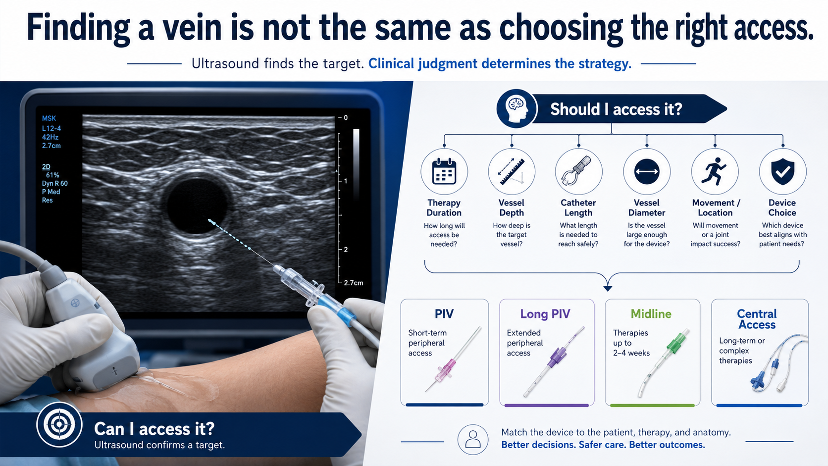
Ultrasound has changed peripheral intravenous access.
It has expanded what clinicians can see, what vessels they can reach, and, in many cases, what they believe they can successfully cannulate. A vein that is invisible and nonpalpable at the skin surface can suddenly become an inviting circular target on a screen. Depth can be measured. Diameter can be estimated. The needle can be followed toward the vessel in real time.
That capability is powerful.
But it also creates a problem.
The ability to identify a target is not the same as having a vascular access strategy.
A vessel may be visible. It may be technically accessible. It may even be successfully cannulated. None of those things, by themselves, establish that it is the right vessel, the right device, or the right access plan for the therapy that patient needs.
And that distinction matters.
Ultrasound Changed the Question
Before ultrasound became widely available for peripheral IV insertion, clinicians were largely limited by what they could see and feel. Difficult access often meant repeated attempts, escalation to another clinician, or consideration of a different vascular access device.
Ultrasound changed those limitations.
Deep veins became accessible. Larger veins became visible. Vessels previously considered outside the practical reach of routine peripheral cannulation could now be targeted.
For patients with difficult intravenous access, this has been an important advance. Ultrasound-guided peripheral IV insertion can reduce failed attempts and provide access when traditional landmark-based techniques have been unsuccessful.
But technology has a tendency to change behavior.
Once clinicians discover that they can reach deeper vessels, the clinical question can quietly shift from:
“What vascular access does this patient need?”
to:
“What vein can I get?”
Those are not the same question.
The first is a clinical assessment.
The second is a technical challenge.
A Target Is Not an Assessment
Ultrasound is exceptionally good at helping clinicians find vessels.
It can help answer questions such as:
Where is the vein?
How deep is it?
What is its approximate diameter?
Is it compressible?
What structures are nearby?
Can I visualize a path for the needle?
All of those observations are useful.
But vascular access assessment requires considerably more.
Before selecting a vessel and inserting a catheter, the clinician should also be asking:
What therapy is prescribed?
How long is that therapy expected to continue?
What are the characteristics of the infusate?
How frequently will the device be used?
Is the patient likely to require repeated blood sampling?
What vascular access has already failed?
What vessels should be preserved for future therapy?
What catheter length will be required to reliably maintain an adequate portion of the catheter within the vessel?
What is the relationship between catheter diameter and vessel diameter?
Where will the catheter tip ultimately reside?
Will the selected site tolerate normal patient movement?
And perhaps most importantly:
Is a peripheral IV actually the most appropriate device for this patient's treatment plan?
Ultrasound can help answer some of these questions.
It cannot replace the clinician who must integrate all of them.
The Seduction of the Big Vein
One of the most compelling ultrasound images is a large, dark, compressible vein sitting several centimeters below the skin.
To an experienced ultrasound user, it can be difficult to resist.
There is the target.
There is the solution.
Or is there?
Deep vessel cannulation introduces considerations that may not be obvious once attention becomes focused on needle placement.
The deeper the vessel, the longer the catheter generally needs to be to maintain adequate intravascular purchase. A short peripheral catheter placed into a deep vessel may initially function beautifully, yet have relatively little catheter remaining within the vessel after traversing the tissue between the skin and vein.
Movement, tissue compression, dressing tension, and changes in arm position can then affect catheter stability.
The insertion may have been technically perfect.
The device selection may still have been poor.
This is where procedural success and clinical success begin to diverge.
Successful Insertion Is Not the Endpoint
Vascular access clinicians often celebrate first-attempt success.
Understandably so.
Repeated needle punctures are painful, inefficient, and frustrating for patients and clinicians alike.
But first-attempt success is only one outcome.
A catheter that is inserted successfully but fails hours later has not solved the patient's vascular access problem. It has postponed it.
A more meaningful definition of success might be:
Can we establish the most appropriate vascular access device, in the most appropriate vessel, with the greatest likelihood of completing the prescribed therapy while minimizing complications and preserving the patient's vasculature?
That is a much higher standard.
And it changes how ultrasound should be used.
Instead of functioning simply as a targeting device, ultrasound becomes part of a broader vascular assessment.
Think Beyond the Insertion Site
One of the greatest limitations of insertion-focused thinking is that the clinician's attention naturally concentrates on the few centimeters surrounding the puncture site.
But the therapy does not end at the puncture site.
Neither does the catheter.
The clinician should consider the entire relationship among skin entry point, vessel depth, catheter length, vessel diameter, catheter tip location, joint movement, and anticipated therapy.
A vein that appears ideal at one point may narrow proximally.
A vessel that looks attractive in the antecubital region may place the catheter in an area exposed to frequent movement.
A deep brachial or basilic vein may be readily cannulated, but the catheter selected may not provide sufficient length for reliable dwell.
A vessel may accommodate the catheter being considered, but another device may better match the duration or nature of therapy.
This is why scanning should not simply identify a vein.
Scanning should help clinicians understand the vascular pathway.
Ultrasound Should Expand Assessment, Not Just Capability
There is an important difference between ultrasound-guided insertion and ultrasound-informed vascular access.
Ultrasound-guided insertion asks:
Can I put a catheter into this vessel?
Ultrasound-informed vascular access asks:
What does this patient's vasculature tell me about the best way to complete therapy?
That second question invites a much broader examination.
The clinician can assess vessel size, depth, course, compressibility, surrounding anatomy, potential insertion zones, and alternative vessels before ever opening a catheter package.
That information can then be combined with the treatment plan.
Perhaps the best answer is a standard peripheral IV.
Perhaps it is a longer peripheral catheter.
Perhaps a midline is more appropriate.
Perhaps the patient's therapy and anticipated duration justify central access.
The purpose of assessment is not to justify the device we already intended to place.
The purpose of assessment is to determine which device makes the most sense.
Difficult Access Does Not Automatically Mean Deep Access
There is another assumption worth challenging.
When a patient is labeled as having difficult intravenous access, clinicians may immediately reach for ultrasound and begin searching for deep upper-arm vessels.
But "difficult access" describes a problem.
It does not prescribe a solution.
The best vessel may still be relatively superficial.
A careful ultrasound survey may reveal vessels that are difficult to visualize externally but remain shallow enough for a conventional-length catheter. In other patients, vessel quality, therapy duration, previous failures, or the vascular anatomy may point toward another device entirely.
The goal should not be to find the deepest vessel we are capable of cannulating.
The goal should be to identify the access option most likely to survive the therapy.
Technology Can Create Procedural Tunnel Vision
Every new clinical technology introduces the possibility of what might be called procedural tunnel vision.
We become increasingly skilled at performing a procedure and gradually begin to view more patients as candidates for that procedure.
Ultrasound is no exception.
The screen commands attention. Needle visualization requires concentration. The challenge of advancing a needle into a small or deep vessel can become absorbing.
But the most important decision may have occurred before the needle ever touched the skin.
Should this vessel have been selected at all?
Highly skilled clinicians sometimes need greater restraint, not greater capability.
The more procedures we know how to perform, the more important it becomes to recognize when not to perform them.
That is part of clinical expertise.
From Vessel Hunting to Vessel Health
Perhaps the greatest opportunity created by ultrasound is not improved needle guidance.
It is improved vascular assessment.
Rather than hunting for a vessel that will accept a catheter, clinicians can begin thinking in terms of vessel health and preservation.
Which vessels are available?
Which should be preserved?
Which provide the best relationship between vessel diameter and catheter size?
Which insertion locations minimize disruption from movement?
What is the likely trajectory of the catheter?
What future vascular access might this patient require?
This changes ultrasound from a rescue technology into a planning technology.
And that may ultimately be its greatest contribution to vascular access practice.
The Better Question
When an ultrasound image presents a beautiful target, the temptation is to think:
I can get that vein.
Perhaps you can.
But expertise requires one additional question:
Should you?
The best vascular access clinicians are not simply those who can cannulate the most difficult vessels.
They are the clinicians who understand when a vessel should be cannulated, when it should be preserved, when another device would be more appropriate, and how today's vascular access decision will affect tomorrow's options.
Ultrasound gives us extraordinary power to see beneath the skin.
What it cannot provide is judgment.
That remains our responsibility.
And ultimately, the goal of vascular access is not to successfully insert a catheter.
The goal is to successfully complete the therapy.